
YOU WANT TO X-RAY MY DOG’S/CAT’S TEETH?
As a vet one of the most common questions I get asked when discussing dental disease and cleanings are “Why does your clinic encourage radiographs and others do not?” and “Can’t you just see which teeth need to be pulled?”. I am certain that almost every technician taking the radiographs and every vet reading them would agree that it would be nice to not need them. Dental radiographs can be difficult to take properly and are time-consuming. Reading these radiographs takes time and experience. BUT they are oh so important!
So here is an example….
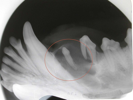
My first day here at Hillcrest, Dr Steen called me into surgery (a neuter in fact) to look at the dog’s mouth. One of the dental arcades was normal, and the other was missing a tooth. Well without dental radiographs we would assume that this dog was just born without that tooth BUT when viewing the radiographs he actually had an unerupted tooth. “So what!” you say? This is really important to know and deal with while he is already under general anesthetic instead of taking the ‘wait and see’ approach.
These retained teeth will cause resorption of the surrounding bone, they then produce fluid in this now open space and form what is called a DENTIGEROUS CYST. This small cyst goes undetected as initially it is non-painful. It continues to produce fluid and grow until it breaks through the bone. Unfortunately, by the time we can detect it there is irreversible damage to the jaw bone and usually to the roots of the teeth near by it. So Thank you, Dr Steen, for detecting this during a routine neuter and dealing with it before it caused this dog permanent damage and the owner a lot of money!
There are also more obvious reasons to take dental radiographs. As a part of a routine dentistry the first thing we do is take x-rays of all the teeth to see if there are any signs of disease below the gumline. Often we can see a ton of tartar but when scaled off the tooth beneath looks beautiful BUT is it really beautiful below the gumline? The x-ray allows us to see the root of the tooth. Even if the exposed tooth looks great we may have to extract that tooth if the roots are diseased. We can also see cavities through the tooth enamel. These teeth are also extracted unless the owner is willing to refer for fillings or a root canal which can be expensive. We know these teeth will cause pain and need to come out at some point so we preemptively remove them.
After extraction, it is equally important to repeat the radiographs to ensure the entire tooth has been successfully removed. I know from personal experience of having a root tip left in my own mouth that there is not much that is more PAINFUL than a portion of root left in the site. It is also a possible source of ongoing future infections. Our governing body the College of Veterinarians of Ontario recommends dental radiology as a professional practice standard when performing veterinary dentistry.
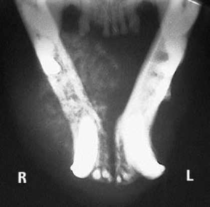
The final reason for dental radiographs is to rule-out bone cancer. Oral tumours are becoming more common in our pets. Is it a tooth root abscess causing swelling and bony changes or is it a bone tumour? A traditional ‘survey’ radiograph using the radiology unit that takes the chest and abdominal rads is often unsuccessful at taking jaw rads. An intraoral dental radiology unit allows us to pinpoint the exact suspect area without underlying structures from the other side of the jaw getting in our way.
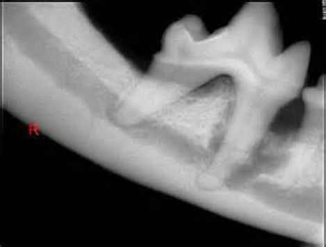
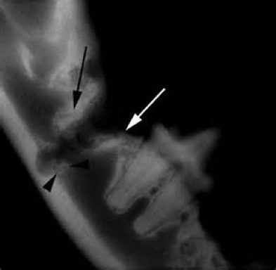
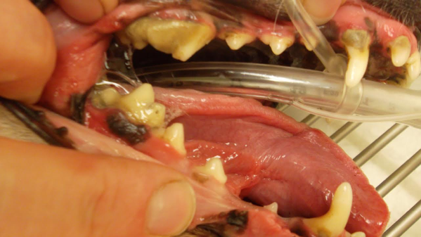
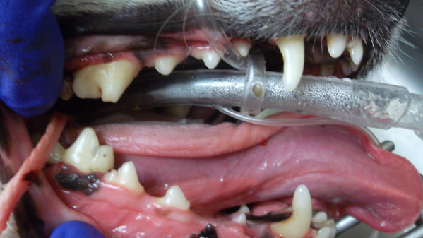
Here is a final case for you to read about. It includes most of the above reasons of why we want dental radiographs. This past Friday I saw a great little dog, Zozo, who was in a lot of pain (if I were him I would have bitten me!). His last 2 physical exams there were notes of significant dental disease. The recommended dental cleaning was not done. The tartar continued to accumulate but even worse he developed a tooth root abscess. On exam, I noted a large bony swelling under the lower jaw on one side and the upper jaw under the eye on the other side. It could have a been a very bad tooth root abscess or bone cancer. Obviously, we needed to know. The dental disease and pain were so bad at this point that we needed to start antibiotics and pain medications. The dental procedure was performed ASAP. The crown of the teeth was actually quite nice when the tartar was removed, but the radiographs tell a different story…
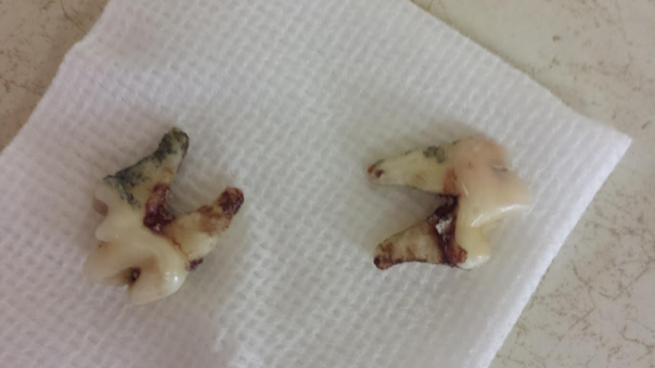
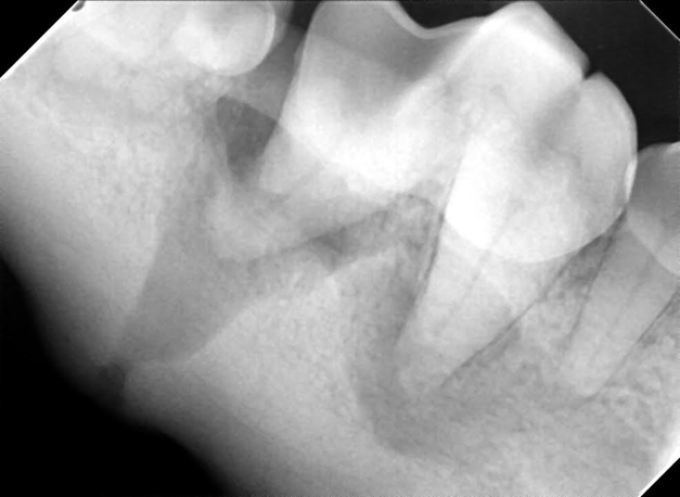
The radiographs revealed a tooth root abscess with a draining tract right through the bone. The jaw is very vulnerable at this location and can easily be fractured. Luckily we knew where the compromised bone was and took extra care. Zozo woke up and was feeling better than ever!
Written by Hillcrest Animal Hospital